
This is the long-form version: six lines of published evidence and the framework that ties them together. Lighter entry points: The Scoliosis Gene and the Nervous System and Not Everyone Has the Gene.
Six lines of published research, from seven different fields, point toward the same place when you read them together: the nervous system. They are a strong lead on a question the orthopedic model has long left open. Why does a straight spine start to curve in a healthy teenager?
Several independent lines of research link adolescent idiopathic scoliosis (AIS) to the nervous system and make it a serious candidate for part of the cause. In mice, losing proprioceptive (position-sense) neurons on both sides produced scoliosis without a prior bone or muscle defect (Blecher et al., 2017). One of the strongest genetic signals for AIS lies near LBX1, a gene that in mice is required for one of the two main classes of dorsal-horn sensory interneurons, which relay pain and touch, some of which handle position signals (Gross et al., 2002; Satoh et al., 2016); whether the human risk acts through those neurons is not confirmed. In people with AIS, position-sense deficits, shifted sensory weighting and altered body perception are documented in subgroups, but they were measured after the curve appeared and could be effects of it. The Neural Generation Hypothesis (Miller, 2026, preprint) proposes that the body schema holds the actively correctable part of the curve; this is untested in people.
The pieces sit in different journals, in fields that rarely read each other.
Each line is peer-reviewed. Each points at the nervous system from a different angle.
My argument in this piece: in adolescent idiopathic scoliosis, the nervous system may help hold part of the curve in place.
Not a single gene acting alone. Not only the bone or the muscles. The body’s internal map, working with less reliable sensory input during a fast-growth window. Others have argued for a nervous system role before, among them Herman in 1985 and Burwell in 2008. What I add is a specific mechanism, built on how the brain predicts the body, and the tests that could prove it wrong.
The full synthesis is a free preprint: The Neural Generation Hypothesis of Adolescent Idiopathic Scoliosis.
Is there evidence that scoliosis originates in the nervous system rather than the spine?
Several lines of research point to the nervous system as a candidate contributor to adolescent idiopathic scoliosis (AIS). Mice lacking proprioceptive neurons develop scoliosis without a prior bone or muscle defect (Blecher et al., 2017). A brain lesion of the body-sensing cortex in developing rats produced scoliosis in 46% of operated animals and none of the controls (Domenech et al., 2013). The LBX1 locus, one of the strongest genetic signals, lies near a gene that builds one of the two main classes of dorsal-horn sensory interneurons in mice (Gross et al., 2002). In people with AIS, studies document position-sense deficits in a subgroup (Lau et al., 2022; 2025), shifted sensory weighting (Simoneau et al., 2006; Pialasse et al., 2015), altered body perception (Picelli et al., 2016; Le Berre et al., 2019) and brain differences (Formaggio et al., 2022; Paramento et al., 2024). These human findings were measured after onset and could be effects of the curve. Whether degraded sensing helps cause AIS in people has not been shown.
Line 1: the genetics point partly at the nervous system
One of the strongest gene signals for scoliosis sits near LBX1. It was found in a genome-wide association study in 2011 and confirmed in a meta-analysis of more than 34,000 people.
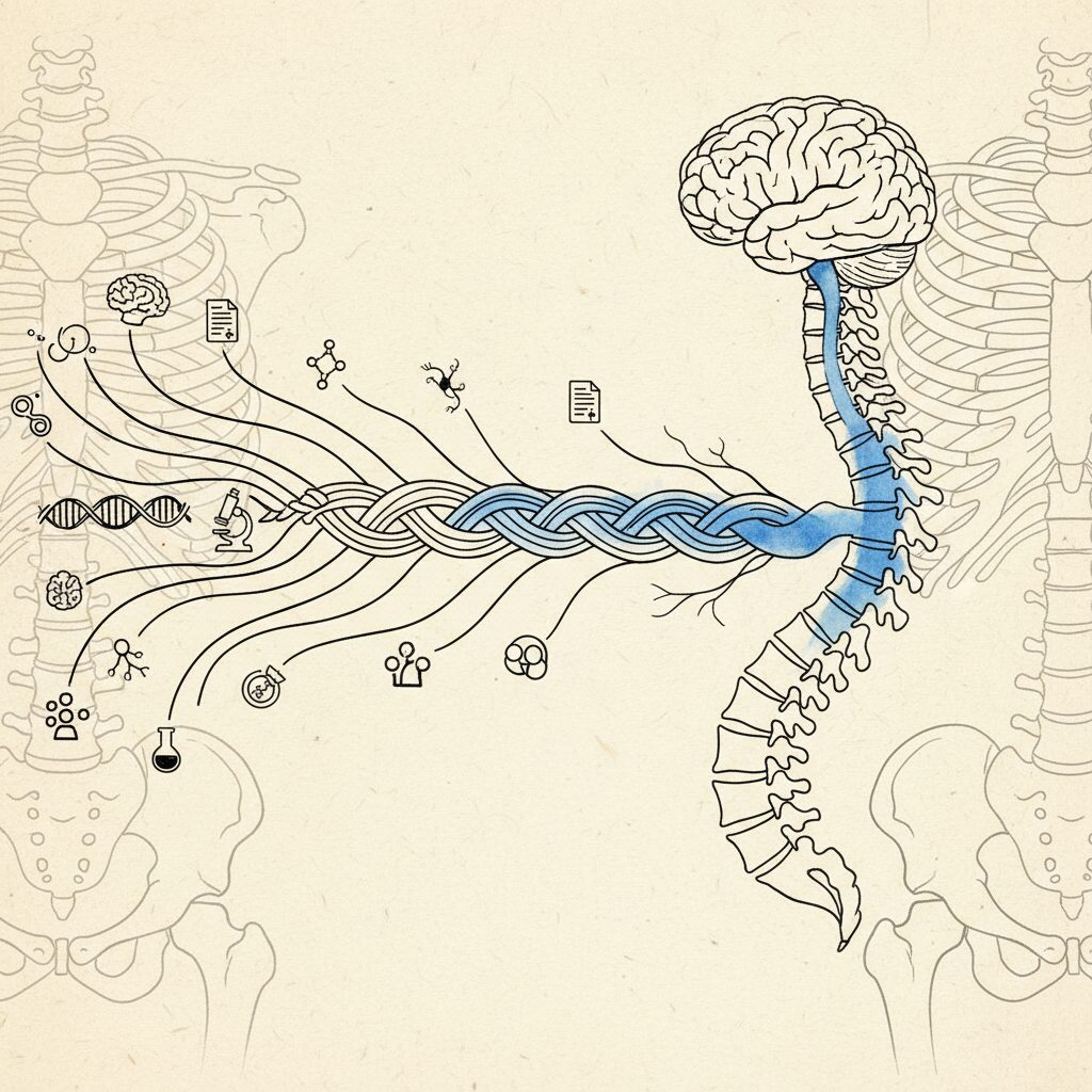
Scoliosis risk genes act on the spine itself and possibly on the nervous system. The LBX1 locus is one of the most replicated genetic signals in adolescent idiopathic scoliosis (Takahashi et al., 2011), with modest effects (pooled odds ratio about 1.2 across 34,626 subjects; Cao et al., 2016). In mice, Lbx1 is required for one of the two major classes of dorsal-horn sensory interneurons, which relay pain and touch, some of which handle position signals within the cord (Gross et al., 2002; Müller et al., 2002; Satoh et al., 2016). Whether AIS risk at this locus acts through these neurons is not confirmed, and LBX1 also guides limb-muscle precursors. A 2024 mouse study (McCallum-Loudeac et al.) deleted a 189 base-pair region near Lbx1: the mice scored worse on a sensorimotor test at 4 weeks and had about 6 degrees of vertebral rotation as adults, but the deficit later converged with normal mice and the deletion also changed neighbouring genes. PAX1 and GPR126 act on the spine’s cartilage and growth plates.
When the media reports this, the framing is: scientists found a scoliosis gene. The implication is that the gene builds curved spines.
It does not.
LBX1 is a transcription factor: a molecular switch that helps decide what kind of neuron a developing cell becomes. In the back part of the spinal cord, where sensory signals first arrive, it separates two main classes of neurons. Mice born without it lose many of the sensory association neurons there. The pathways affected are the ones that relay pain and touch, and some of these cells also handle signals about body position. LBX1 also guides muscle precursor cells into the limbs, so it is not a pure sensing gene either.
Other well-replicated genes, PAX1 and GPR126, shape the spine’s own cartilage and growth plates. So the genetics point two ways: at the structure, and at the sensing.
A 2024 mouse study went after the LBX1 region directly. Mice missing a small stretch of DNA nearby did worse on a sensorimotor test at four weeks old. As adults, their vertebrae were rotated by about six degrees.
Striking. And it is one study. The deficit faded later, the deletion changed other genes too, and whether the human gene works through sensing is still open. But it is a strange place to find a spine gene, and it is where this line of thinking started.
Published: Takahashi et al., 2011, Nature Genetics. Cao et al., 2016, BMC Musculoskeletal Disorders. Gross et al., 2002, Neuron. McCallum-Loudeac et al., 2024, Human Molecular Genetics.
Line 2: losing position sense can start a curve, in animals
This is the strongest causal evidence, and most people have never heard of it.
In 2017, a research team studied mice born without proprioceptive neurons, the sensors that report where the body is. By about three months old, 19 of 20 had scoliosis. Among 20 normal littermates, one had a small curve. In mice followed by scans, the spines were straight at 40 days and curved by 60, around puberty, and their vertebrae and back muscles looked normal before the curves appeared. Removing the same gene only in bone, cartilage or tendon did nothing. Removing it only in the sensory neurons curved the spine in about 40% of the mice.
Older one-sided experiments also show that sensing matters, though they produce a nerve-and-muscle type of curve. Cutting sensory nerve roots on one side in monkeys produced scoliosis, and the more roots cut, the bigger the curve. In young rats, removing part of the brain’s body-sensing region produced scoliosis in almost half of them, and in none of the comparison rats.
People have their own natural experiments. Rare conditions that remove or reduce position sense, such as familial dysautonomia and PIEZO2 deficiency, often come with scoliosis, though they bring other problems too, such as weakness or autonomic failure.
These are big losses, not the subtle differences seen in ordinary teenagers, and the mice without position sensors were also badly uncoordinated. What they show is that sensing matters for keeping a spine straight.
In animals, yes. Mice lacking proprioceptive neurons (Runx3 knockout) developed scoliosis in 19 of 20 cases by postnatal day 90, against 1 of 20 littermates, without preceding differences in vertebral shape or paraspinal muscle; deleting the gene only in bone, cartilage or tendon produced no deformity (Blecher et al., 2017). Dorsal root section in monkeys produced scoliosis convex to the cut side, with severity dependent on the number of roots cut (Pincott et al., 1984). A right somatosensory cortex lesion in developing rats produced scoliosis in 46% of operated animals and none of the sham or intact controls (Domenech et al., 2013; one group, not replicated). In humans, conditions with severe proprioceptive loss, such as familial dysautonomia (Hayek et al., 2000; Macefield et al., 2024) and biallelic PIEZO2 deficiency (Chesler et al., 2016), carry high rates of scoliosis but involve other deficits. Whether the milder sensory differences seen in adolescent idiopathic scoliosis can start a curve is untested.
Published: Blecher et al., 2017, Developmental Cell. Pincott et al., 1984, Journal of Bone and Joint Surgery. Domenech et al., 2013, The Spine Journal.
Line 3: position sense is weaker in a subgroup
Proprioceptive deficits in scoliosis have been reported for over two decades. A 2022 meta-analysis found that teenagers with AIS make larger errors when asked to return a joint to a position, and need a bigger movement before they notice it. In a 2025 study that tested the trunk directly, about 30% of patients fell below normal values, and those patients had larger curves. In a one-year follow-up, that trunk deficit did not predict which curves progressed.
One common reading is that these deficits are a result of the curve: the spine bends, the body map gets distorted, position sense suffers. That is a reasonable reading, and the data cannot rule it out. Every one of these measurements was taken after the curve had appeared. A 2014 systematic review made the same point: the abnormalities “were never studied in AIS cases before the onset of the deformity.”
One finding stands out. Assaiante and colleagues reported in 2012 that adolescents in general lean less on position sense while they grow, and that in scoliosis the static sense of position looks normal while the dynamic sense, the one that tracks movement, appears affected. My read: during the growth years, the channel that would notice slow drift may be the one getting the least attention. That is a proposal, from a study of 10 patients.
Do proprioceptive deficits cause scoliosis or result from it?
In people, this is not yet known. A 2022 meta-analysis of 11 case-control studies found larger repositioning errors (about 1.27 degrees, pooled from three studies) and higher motion-detection thresholds (about 1.6 degrees, pooled from two) in adolescent idiopathic scoliosis, measured at the neck, elbow and knee rather than the trunk (Lau et al., 2022). About 30% of patients had a trunk deficit, associated with larger curves (Lau et al., 2025), but in a one-year follow-up the trunk deficit did not predict progression (Lau et al., 2026). A systematic review noted that these abnormalities were never studied before onset and may result from the deformity (Schlösser et al., 2014). In animals, loss of proprioception can cause scoliosis (Blecher et al., 2017). The Neural Generation Hypothesis (Miller, 2026) proposes a causal role in some adolescents and lists pre-onset tests; it is untested.
Published: Lau et al., 2022, Global Spine Journal. Lau et al., 2025, Bone & Joint Open. Lau et al., 2026, North American Spine Society Journal. Schlösser et al., 2014, PLoS One. Assaiante et al., 2012, PLoS One.
Line 4: sensory weighting shifts toward the inner ear
Your brain does not treat all sensory channels equally. It gives each one weight according to how reliable it seems. When one channel gets noisy, it leans on the others. That is Bayesian inference applied to balance, and it is how the system stays upright when one input drifts.
Simoneau’s research group in Quebec has studied this in scoliosis for years. In a small 2006 study (8 patients), they found that adolescents with scoliosis have trouble re-balancing their senses after a disturbance. Their later model-based estimates put the weight teenagers with AIS give to the inner ear at about 13%, against about 6% in healthy teenagers. Roughly double. And the same in mild and severe curves.
That is what you would expect if position sense were trusted less. The inner ear tells you which way is down. It is not built to report fine trunk position. A body leaning on it more may run on a coarser map: good enough to stand, maybe not precise enough to catch a slow drift during growth. That last step is a proposal, not a measurement.
Two cautions. These are estimates from a model, and much of this work comes from one research network. In a larger sample, the same group found impaired sensorimotor control in 42.5% of patients, not all of them. And other studies point the other way (more on that below).
How does sensory reweighting contribute to scoliosis?
The brain weights each sensory channel by its estimated reliability (Friston, 2010). Simoneau et al. (2006) found that adolescents with idiopathic scoliosis have difficulty reweighting sensory inputs after a perturbation. Using galvanic vestibular stimulation and a neuromechanical model, Pialasse et al. (2015) estimated that they assign about 13% weight to vestibular input versus about 6% in controls, with the same weighting in mild and severe curves. These are model-derived estimates, largely from one research network, and a larger study found impaired sensorimotor control in 42.5% of patients (Pialasse et al., 2017). A systematic review instead reported greater reliance on visual and proprioceptive information (Paramento et al., 2024); the measures differ and the direction of reweighting is not settled. The Neural Generation Hypothesis proposes, untested, that heavier reliance on vestibular input leaves slow trunk drift during growth below the correction threshold.
Published: Simoneau et al., 2006, BMC Neuroscience. Pialasse et al., 2015 and 2017, Gait & Posture.
Line 5: the body’s sense of its own alignment is altered
The body schema is the brain’s internal model of the body in space. Not the conscious image you have of how you look. The non-conscious, always-running model that guides movement. Where your shoulders sit. How your ribcage rotates. What your pelvis does when you walk.
The concept is over a century old. Head and Holmes described it in 1911. Paillard sharpened the line between body schema and body image in 1999.
In 2016, Picelli and colleagues asked directly: do adolescents with idiopathic scoliosis have body schema disorders? They asked 44 teenagers with scoliosis to match their own trunk to pictures. The teenagers misjudged their trunk alignment in patterns specific to their curve type, overestimating in some areas and underestimating in others. The study had no control group.
Another group measured the felt sense of upright. In two studies, adolescents with scoliosis felt “straight up” about two degrees toward the outer side of their curve, while their visual sense of vertical was normal. A third study found patients were more sensitive than controls to curvature in pictures of spines, not less.
So the body’s sense of itself is altered, but not simply blind to the curve. A small shift in felt upright, mixed judgments of shape, and sharper attention to curvature.
And here is the number that should keep the research community up at night. In 2022, Bertuccelli and colleagues reviewed every study of body image and body schema in teenagers with idiopathic scoliosis published between 2000 and 2021. They found 27. Twenty-four looked at body image: how teenagers feel about how they look. Four looked at body schema.
Four out of twenty-seven (some studies looked at both).
Same word, “body.” Completely different question. One asks how the condition feels. The other asks what the brain’s model of the body is doing. The field has mostly asked the first.
What is the body schema and how does it relate to scoliosis?
The body schema is the brain’s non-conscious internal model of the body in space, which guides posture and movement (Head and Holmes, 1911; Paillard, 1999). In adolescent idiopathic scoliosis, Picelli et al. (2016) found that 44 patients (no control group) misjudged their own trunk alignment in curve-type-specific patterns. Le Berre et al. (2019) and Catanzariti et al. (2022) found the subjective postural vertical shifted about 2 degrees toward the convex side, with a normal visual vertical. Patients also detected spinal curvature in images at lower thresholds than controls (Virovec et al., 2026). Bertuccelli et al. (2022) reviewed 27 studies on body representation in AIS: 24 assessed body image and only 4 assessed body schema. All of these measurements were taken after onset.
Published: Picelli et al., 2016, Journal of Back and Musculoskeletal Rehabilitation. Le Berre et al., 2019 and Catanzariti et al., 2022, Spine Deformity. Virovec et al., 2026, Experimental Brain Research. Bertuccelli et al., 2022, Adolescent Research Review. Head & Holmes, 1911, Brain. Paillard, 1999.
Line 6: brain, balance and inner ear show differences
If sensing works differently, the brain should show it. Several studies suggest it does.
In 2022, researchers measured brain rhythms in 14 adolescents with scoliosis and 14 without. The scoliosis group showed more theta activity over central areas and lopsided alpha activity over the sensorimotor cortex, the strip of brain that maps the body.
In 2024, another team used machine learning on brain scans to tell 32 patients with severe scoliosis apart from 31 controls. With that many brain regions measured in that few people, the result needs repeating, and severe curves cannot tell us what came first.
A 2024 systematic review of 61 studies found that teenagers with AIS show poorer standing balance and lean more on vision and position sense. That sits awkwardly with the inner-ear findings above; the studies used different measures, and the field has not settled which way the weighting goes. The review called for more work on the nervous system’s role in how scoliosis develops.
The inner ear story is mixed. A 2015 review found clinical support for a vestibular role, while noting it has not shown up in every study. Two small scan studies found the balance canals of the inner ear shaped differently in scoliosis, which the authors argue is probably set before birth, though they disagree on the direction. In chickens whose pineal gland was removed, a long-used model, the authors suggested melatonin loss disturbs the growth of the position-sense system; the same surgery produced no scoliosis in monkeys.
None of these studies can say whether the differences came before the curve or after it.
Brain and balance differences are documented in adolescent idiopathic scoliosis, but their direction is unknown. Formaggio et al. (2022; 14 patients, 14 controls) found increased theta power over central areas and lateralized alpha activity over sensorimotor cortex, with no balance difference between the groups in that sample. Payas et al. (2024) classified 32 patients with severe AIS against 31 controls from brain volumetric measures; overfitting is a concern and severe curves cannot date the change. A systematic review of 61 studies (Paramento et al., 2024) found impaired standing balance and greater reliance on visual and proprioceptive information, which sits uneasily with model estimates of heavier vestibular weighting (Pialasse et al., 2015); the measures differ and the direction of reweighting is not settled. The review did not conclude that brain changes are causes rather than consequences. Vestibular findings are mixed (Hawasli et al., 2015; Hitier et al., 2015; Carry et al., 2020).
Published: Formaggio et al., 2022, Scientific Reports. Payas et al., 2024, JOR Spine. Paramento et al., 2024, PLoS One. Hawasli et al., 2015, European Spine Journal. Hitier et al., 2015 and Carry et al., 2020, PLoS One. Machida et al., 1995, Journal of Bone and Joint Surgery (British). Cheung et al., 2005, Spine.
The framework: how a predicting brain could hold a curve
The framework I use to connect the six lines was set out by Karl Friston in a 2010 review.
His free-energy principle, and the active inference framework built on it, describe the brain as a prediction engine. Not a reactor. A predictor. It keeps a running model of the body and the world and checks incoming signals against it. Signals it trusts change the model. Signals it does not trust get less say.
Apply that to a growing spine, and here is what the Neural Generation Hypothesis proposes. During the growth spurt the spine drifts a tiny amount each month. With clear body sense, the nervous system notices and corrects it. With blurry body sense, the drift is too small to register as change, so nothing corrects it. Meanwhile the map keeps quietly updating to match the body it is in. Month by month, the curve becomes the map’s new “straight,” and the body is held there.
The map is not too slow. It updates toward the curve instead of back to straight.
Two limits. The hypothesis claims only the part of the curve a person can actively correct, beyond what simple flexibility allows. How big a curve grows is driven by uneven loading on growing bone, which is well established and not part of the claim.
How does the predictive coding framework explain scoliosis?
Under active inference (Friston, 2010), the brain keeps a predictive model of the body, the body schema, and weights each sensory signal by its estimated precision. The Neural Generation Hypothesis (Miller, 2026; preprint) applies this to adolescent idiopathic scoliosis. It proposes that where the sense of trunk position is degraded, drift during the growth spurt is too slow to be signalled as change, so it is not corrected, and the body schema’s slow learning moves its reference toward the curve until the curve is treated as straight. The claim covers only the actively correctable part of the curve; growth-driven asymmetric loading sets curve magnitude. Each step specific to the hypothesis is untested, and the paper lists eleven testable predictions.
The proposed chain
Put together, the hypothesis proposes a chain. The tag on each step says where the evidence stands.
- Risk genes set a small liability, partly on the sensing side (LBX1) and partly in the spine itself (PAX1, GPR126). Supported as risk; the sensing route is unconfirmed.
- Position sense is less precise in some adolescents. Seen in a subgroup, measured after the curve appeared.
- The brain leans more on other channels, such as the inner ear. Model-based estimates, measured after the curve appeared; other studies disagree on the direction.
- During the growth spurt, slow drift goes unnoticed. Untested.
- The map updates toward the curve and holds the body there. Untested.
- The curve, and the stress around it, feed back and help it stick. The position-sense link is an association; the stress and social loops are untested.
Each step on its own has been studied, in a different field. The chain as a whole is a proposal, with written tests that could prove it wrong. That is what the preprint adds: read the full paper here.
What’s shown, and what isn’t
- Shown: scoliosis risk genes exist, and uneven loading during growth drives how big curves get.
- Shown in animals: losing position sense entirely can start a curve. Untested in teenagers with AIS.
- Seen in people, but only after the curve appeared: weaker position sense, shifted sensory weighting, an altered sense of upright and brain differences, each in some patients. These could be a result of the curve rather than its cause.
- Suggestive: the LBX1 mouse study. The deficit faded later, and the deletion also changed nearby genes.
- Not yet tested: the steps that belong to this hypothesis. Drift too slow to notice, the map updating toward the curve, the stress and social loops, and whether the held part can change.
The paper rates every link this way. Two tests could move it soon: balance data already collected on thousands of 10-year-olds, checked against scoliosis at 15, and perception measured when a brace comes off.
What this changes
If this chain holds up, three things follow.
First: “idiopathic” may name a measurement gap. Most scoliosis that appears in childhood or adolescence carries that label because no cause has been established. This hypothesis is one candidate explanation for part of it. In my view the cause is not unknowable. It has been under-measured. Current tools measure the output. The proposed mechanism lives in the system generating the output.
Second: “genetic” does not mean “fixed.” The genes set a small risk. If the hypothesis is right, part of what holds the curve is a map the brain keeps updating, now, in real time. A map that updates is, at least in principle, a map that can take in new information. Whether that changes a curve is one of the things the paper says still has to be tested.
Third: the questions change. If scoliosis is only a bone problem, you brace it, strengthen around it, or fuse it. If part of it is held by a map running on blurry input, you also ask what the map is using, and whether better input could help. That does not replace monitoring, bracing or surgery. It is another address to look at.
Instead of only asking how to correct the curve, the question becomes: what is the body schema using to hold this curve, and what would it take to give it better data?
What would change if scoliosis were treated as a nervous system problem?
If part of adolescent idiopathic scoliosis is held by the body schema, assessment would add measures of the sensing side: proprioceptive testing, sensory-weighting evaluation (Simoneau et al., 2006; Pialasse et al., 2015) and body schema or perceived-vertical tests (Picelli et al., 2016; Le Berre et al., 2019). The Cobb angle would remain the structural measure. The Neural Generation Hypothesis (Miller, 2026) makes no claim that any intervention reverses established deformity or displaces monitoring, bracing or surgery. Existing scoliosis-specific exercise methods already include sensorimotor elements; no trial has yet shown that changing perception changes the curve, and the paper proposes how to test it.
The emerging picture
This is not one study making a bold claim. It is six lines of research, across four decades and several countries, each showing one piece of a picture that only comes into view when you read them together.
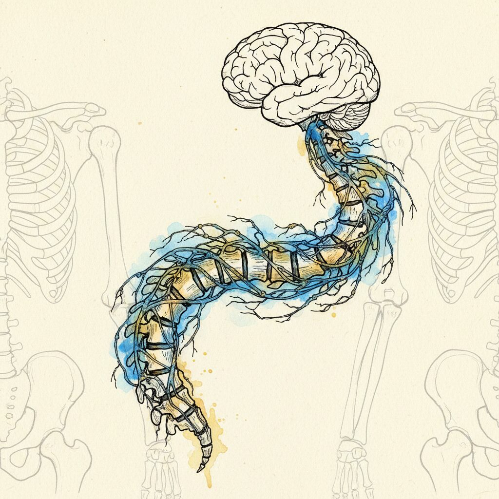
The genetics point partly at the nervous system. The experiments show that losing position sense can start a curve, in animals. The position-sense research, the sensory-weighting research, the body schema research and the brain imaging all find differences in teenagers with AIS. Predictive coding offers a way they could fit together.
What is missing is the test. We found no study that measured position sense in children before a curve appeared and then followed them. That is the study that would test which came first, and the preprint spells out how to run it.
People with AIS are told their curve has no known cause. Part of what I think answers that question is published across journals in fields that rarely read each other. Not because anyone is negligent. Because the model most clinicians are trained in does not look at the nervous system first.
In this view, the spine is the printout. The full synthesis, a hypothesis and not yet a tested finding, is free to read: The Neural Generation Hypothesis (Zenodo preprint).
What medicine calls scoliosis, we read as a pattern the nervous system may be helping to hold. That is a hypothesis, built from published evidence, with the tests written down.
Start of the Generative Posture Series: Why 80% of Scoliosis Cases Have No Explanation (G-1)
Related: The Scoliosis Gene That Points to the Nervous System | How Your Brain Controls Posture | Your Diagnosis Describes a Shape, Not a Cause
Ready to work with the system that generates your posture? The Syntropic Core method addresses posture at the body schema level. Not the shape. The prediction that helps hold the shape. Learn more at syntropiccore.com.
Sources
- Miller, S.A. (2026). The Neural Generation Hypothesis of Adolescent Idiopathic Scoliosis. Version 3.0. Preprint, Zenodo. doi:10.5281/zenodo.19342098. [T3]
Not peer reviewed. States the evidence status of each link and eleven testable predictions. - Takahashi, Y., Kou, I., Takahashi, A., et al. (2011). A genome-wide association study identifies common variants near LBX1 associated with adolescent idiopathic scoliosis. Nature Genetics, 43(12), 1237-1240. [T1]
Original genome-wide association study identifying the LBX1 locus in AIS. - Cao, Y., Min, J., Zhang, Q., Li, H., Li, H. (2016). Associations of LBX1 gene and adolescent idiopathic scoliosis susceptibility: a meta-analysis based on 34,626 subjects. BMC Musculoskeletal Disorders, 17, 309. [T1]
Meta-analysis confirming the LBX1 association, with modest effect sizes. - Gross, M.K., Dottori, M., Goulding, M. (2002). Lbx1 specifies somatosensory association interneurons in the dorsal spinal cord. Neuron, 34(4), 535-549. [T1]
In Lbx1-null mice, dorsal horn association interneurons are lost and sensory innervation of the dorsal horn is disrupted; the affected pathways relay pain and touch. - Müller, T., Brohmann, H., Pierani, A., Heppenstall, P.A., Lewin, G.R., Jessell, T.M., Birchmeier, C. (2002). The homeodomain factor Lbx1 distinguishes two major programs of neuronal differentiation in the dorsal spinal cord. Neuron, 34(4), 551-562. [T1]
Lbx1 separates two major classes of dorsal horn neurons; in mutants, class B neurons take on class A identity. - Satoh, D., Pudenz, C., Arber, S. (2016). Context-dependent gait choice elicited by EphA4 mutation in Lbx1 spinal interneurons. Neuron, 89(5), 1046-1058. [T1]
Some Lbx1-lineage spinal interneurons take part in proprioceptive and locomotor circuits. - McCallum-Loudeac, J., Moody, E., Williams, J., Johnstone, G., Sircombe, K.J., Clarkson, A.N., Wilson, M.J. (2024). Deletion of a conserved genomic region associated with adolescent idiopathic scoliosis leads to vertebral rotation in mice. Human Molecular Genetics, 33(9), 787-801. [T1]
CRISPR deletion of a 189 bp region near Lbx1. Sensorimotor deficit at 4 weeks; about 6 degrees of vertebral rotation at T6 in adults. The deficit converged by 20 weeks, and neighbouring genes also changed. - Blecher, R., Krief, S., Galili, T., Biton, I.E., Stern, T., Assaraf, E., et al. (2017). The Proprioceptive System Masterminds Spinal Alignment: Insight into the Mechanism of Scoliosis. Developmental Cell, 42(4), 388-399. [T1]
Mice lacking proprioceptive neurons: scoliosis in 19 of 20 by day 90, without preceding bone or muscle differences. The mice are also ataxic. - Pincott, J.R., Davies, J.S., Taffs, L.F. (1984). Scoliosis caused by section of dorsal spinal nerve roots. Journal of Bone and Joint Surgery (British), 66(1), 27-29. [T1]
Monkeys: dorsal root section produced scoliosis convex to the cut side; severity rose with the number of roots cut. - Domenech, J., Barrios, C., Tormos, J.M., Pascual-Leone, A. (2013). Somatosensory cortectomy induces motor cortical hyperexcitability and scoliosis: an experimental study in developing rats. The Spine Journal, 13(8), 938-946. [T1]
Right somatosensory cortex lesion: scoliosis in 46% of operated rats, none of the controls. One group, not replicated. - Hayek, S., Laplaza, F.J., Axelrod, F.B., Burke, S.W. (2000). Spinal deformity in familial dysautonomia. Prevalence, and results of bracing. Journal of Bone and Joint Surgery (American), 82(11), 1558-1562. [T1]
Scoliosis common in a condition with poor proprioception; other deficits are also present. - Macefield, V.G., Smith, L.J., Norcliffe-Kaufmann, L., Palma, J.A., Kaufmann, H. (2024). Sensorimotor control in the congenital absence of functional muscle spindles. Experimental Physiology, 109(1), 27-34. [T1]
Familial dysautonomia: congenital absence of functional muscle spindles, with impaired sensorimotor control. - Chesler, A.T., Szczot, M., Bharucha-Goebel, D., et al. (2016). The Role of PIEZO2 in Human Mechanosensation. New England Journal of Medicine, 375(14), 1355-1364. [T1]
PIEZO2 loss: profoundly decreased proprioception with progressive scoliosis, alongside joint and muscle involvement. - Lau, K.K.L., Law, K.K.P., Kwan, K.Y.H., Cheung, J.P.Y., Cheung, K.M.C., Wong, A.Y.L. (2022). Timely Revisit of Proprioceptive Deficits in Adolescent Idiopathic Scoliosis: A Systematic Review and Meta-Analysis. Global Spine Journal, 12(8), 1852-1861. [T1]
11 case-control studies. Pooled values come from neck, elbow and knee joints; case-control designs cannot show cause. - Lau, K.K.L., Kwan, K.Y.H., Cheung, J.P.Y., Law, K.K.P., Wong, A.Y.L., Chow, D.H.K., et al. (2025). A substantial proportion of subjects with adolescent idiopathic scoliosis display spinal and peripheral proprioceptive deficits. Bone & Joint Open, 6(10), 1164-1170. [T1]
166 patients: 30.1% had a trunk proprioceptive deficit, associated with larger curves. - Lau, K.K.L., Kwan, K.Y.H., Cheung, J.P.Y., Law, K.K.P., Wong, A.Y.L., Chow, D.H.K., et al. (2026). Association of proprioceptive deficits with curve progression and treatment outcomes in subjects with adolescent idiopathic scoliosis: a prospective cohort study with 1 year of follow-up. North American Spine Society Journal, 27, 100899. [T1]
The trunk deficit did not predict curve progression over one year of conservative care. - Schlösser, T.P., van der Heijden, G.J., Versteeg, A.L., Castelein, R.M. (2014). How ‘idiopathic’ is adolescent idiopathic scoliosis? A systematic review on associated abnormalities. PLoS One, 9(5), e97461. [T1]
Associated abnormalities were never studied before onset; some may be a result rather than a cause of the deformity. - Assaiante, C., Mallau, S., Jouve, J.L., Bollini, G., Vaugoyeau, M. (2012). Do adolescent idiopathic scoliosis (AIS) neglect proprioceptive information in sensory integration of postural control? PLoS One, 7(7), e40646. [T1]
10 patients, 10 controls. Static proprioception unaffected; the authors conclude dynamic proprioception is mainly affected. - Simoneau, M., Mercier, P., Blouin, J., Allard, P., Teasdale, N. (2006). Altered sensory-weighting mechanisms is observed in adolescents with idiopathic scoliosis. BMC Neuroscience, 7, 68. [T1]
Adolescents with scoliosis had difficulty reweighting sensory inputs after a perturbation. - Pialasse, J.P., Descarreaux, M., Mercier, P., Simoneau, M. (2015). Sensory reweighting is altered in adolescent patients with scoliosis: evidence from a neuromechanical model. Gait & Posture, 42(4), 558-563. [T1]
Model-based estimate: about 13% vestibular weighting in AIS versus about 6% in controls, the same in mild and severe curves. - Pialasse, J.P., Mercier, P., Descarreaux, M., Simoneau, M. (2017). A procedure to detect abnormal sensorimotor control in adolescents with idiopathic scoliosis. Gait & Posture, 57, 124-129. [T1]
Impaired sensorimotor control in 42.5% of patients. - Picelli, A., Negrini, S., Zenorini, A., Iosa, M., Paolucci, S., Smania, N. (2016). Do adolescents with idiopathic scoliosis have body schema disorders? A cross-sectional study. Journal of Back and Musculoskeletal Rehabilitation, 29(1), 89-96. [T1]
44 patients (no control group) misperceived their trunk alignment in curve-type-specific patterns. - Le Berre, M., Pradeau, C., Brouillard, A., Coget, M., Massot, C., Catanzariti, J.F. (2019). Do Adolescents With Idiopathic Scoliosis Have an Erroneous Perception of the Gravitational Vertical? Spine Deformity, 7(1), 71-79. [T1]
Subjective postural vertical shifted about 2 degrees toward the convex side; visual vertical normal. - Catanzariti, J.F., Coget, M., Brouillard, A. (2022). A perception bias of the gravitational vertical is confirmed in Adolescent Idiopathic Scoliosis. Spine Deformity, 10(1), 69-78. [T1]
Second study of the shifted postural vertical, in left lumbar curves. - Virovec, M., Barumerli, R., Cesari, P. (2026). Motor cortex excitability during spine shape-judgment in adolescent idiopathic scoliosis: a TMS motor evoked potential study. Experimental Brain Research, 244(8), 147. [T1]
Patients detected spinal curvature in images at lower thresholds than controls. - Bertuccelli, M., Cantele, F., Masiero, S. (2022). Body Image and Body Schema in Adolescents with Idiopathic Scoliosis: A Scoping Review. Adolescent Research Review, 8, 97-115. [T1]
Of 27 studies, 24 assessed body image and only 4 assessed body schema. - Formaggio, E., Bertuccelli, M., Rubega, M., Di Marco, R., Cantele, F., Gottardello, F., De Giuseppe, M., Masiero, S. (2022). Brain oscillatory activity in adolescent idiopathic scoliosis. Scientific Reports, 12, 17266. [T1]
14 patients, 14 controls: increased theta power and lateralized alpha activity over sensorimotor cortex. - Payas, A., Kocaman, H., Yildirim, H., Batin, S. (2024). Prediction of adolescent idiopathic scoliosis with machine learning algorithms using brain volumetric measurements. JOR Spine, 7(3), e1355. [T1]
32 severe AIS patients versus 31 controls; small sample for the number of brain regions, so overfitting is a concern. - Paramento, M., Passarotto, E., Maccarone, M.C., Agostini, M., Contessa, P., Rubega, M., Formaggio, E., Masiero, S. (2024). Neurophysiological, balance and motion evidence in adolescent idiopathic scoliosis: a systematic review. PLoS One, 19(5), e0303086. [T1]
61 studies: impaired standing balance and greater reliance on visual and proprioceptive information; does not conclude brain changes are causes. - Hawasli, A.H., Hullar, T.E., Dorward, I.G. (2015). Idiopathic scoliosis and the vestibular system. European Spine Journal, 24(2), 227-233. [T2]
Review: clinical and limited animal support for a vestibular role, not reproduced in all studies. - Hitier, M., Hamon, M., Denise, P., Lacoudre, J., Thenint, M.A., Mallet, J.F., et al. (2015). Lateral Semicircular Canal Asymmetry in Idiopathic Scoliosis: An Early Link between Biomechanical, Hormonal and Neurosensory Theories? PLoS One, 10(7), e0131120. [T2]
Inner-ear canal shape differs in AIS in a small MRI study. - Carry, P.M., Duke, V.R., Brazell, C.J., Stence, N., Scholes, M., Rousie, D.L., Hadley Miller, N. (2020). Lateral semi-circular canal asymmetry in females with idiopathic scoliosis. PLoS One, 15(4), e0232417. [T2]
A second small MRI study of inner-ear canal asymmetry in AIS; the direction of asymmetry differs from Hitier et al. - Machida, M., Dubousset, J., Imamura, Y., Iwaya, T., Yamada, T., Kimura, J. (1995). Role of melatonin deficiency in the development of scoliosis in pinealectomised chickens. Journal of Bone and Joint Surgery (British), 77(1), 134-138. [T2]
Pinealectomised chickens developed scoliosis; the authors attributed it to disturbed growth of the proprioceptive system. - Cheung, K.M., Wang, T., Poon, A.M., Carl, A., Tranmer, B., Hu, Y., Luk, K.D., Leong, J.C. (2005). The effect of pinealectomy on scoliosis development in young nonhuman primates. Spine, 30(18), 2009-2013. [T2]
Pinealectomy produced no scoliosis in young rhesus monkeys. - Herman, R., Mixon, J., Fisher, A., Maulucci, R., Stuyck, J. (1985). Idiopathic scoliosis and the central nervous system: a motor control problem. Spine, 10(1), 1-14. [T1]
An early account of AIS as a motor control problem. - Burwell, R.G., Dangerfield, P.H., Freeman, B.J. (2008). Concepts on the pathogenesis of adolescent idiopathic scoliosis. Studies in Health Technology and Informatics, 135, 3-52. [T1]
Includes a body-schema concept within a broader account of AIS pathogenesis. - Paillard, J. (1999). Body Schema and Body Image: A Double Dissociation in Deafferented Patients. Motor Control, Today and Tomorrow, 197-214. [T1]
Body schema as a non-conscious model guiding motor output from sensory input. - Friston, K. (2010). The free-energy principle: a unified brain theory? Nature Reviews Neuroscience, 11(2), 127-138. [T1]
Active inference framework. Sensory precision weighting shapes the generative model’s output. - Head, H., Holmes, G. (1911). Sensory disturbances from cerebral lesions. Brain, 34(2-3), 102-254. [T1]
Original body schema concept. The brain maintains a postural model updated by sensory input.
The Posture Dojo is a free community exploring the science of how your body generates posture. No corrections. No exercises. A different operating system. Join the conversation here.
Related research
- Two Papers, One Framework: The Neural Generation Hypothesis and the Generative Posture Framework
- Stand Up Tall, Pull Your Shoulders Back, Brace Your Core: Why the 3 Most Common Posture Cues Are Neuroscience Dead Ends
- How Much Can I Actually Change My Scoliosis? What the Research Shows
- You Can’t Tickle Yourself. That’s Why Your Posture Won’t Change.
Comments
One response to “The Scoliosis Research That Points to the Nervous System”
[…] The Scoliosis Research That Points to the Nervous System […]